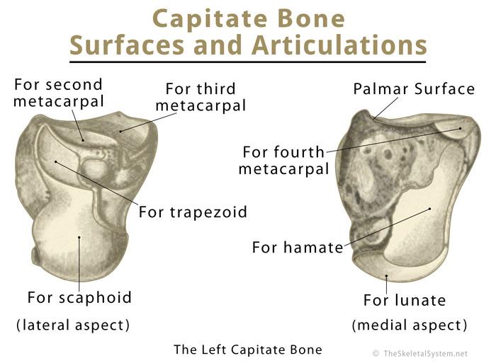
Capitate the capitate is a larger carpal bone that articulates distally with the metacarpal 3 2 and sometimes 4.
Carpal bone siding.
Click on the tags below to find other quizzes on the same subject.
Touch your thumb to every finger like you are.
In human anatomy the main role of the wrist is to facilitate effective positioning of the hand and powerful use of the extensors and flexors of the forearm and the mobility.
The tight space between this fibrous band and the wrist bone is called the carpal tunnel.
Dorsal surface triquetrum 3.
Collectively the carpal bones form an arch in the coronal plane.
This is a key aid in the diagnosing the hamate carpal.
Smallest carpal bone last to ossify sits at a plane anterior to the other carpal bones medial to guyon s canal 2.
While this may make me a bad anatomist editor s note.
The end is squared off whilst the proximal end is rounded.
It does make her a bad anatomist when i m working with whole carpals i rarely rely on their anatomical relationships but instead use cheap tricks to identify and side them.
This is an online quiz called carpal bones.
Palmar surface flexor carpi ulnaris abductor.
Repeat this for five to 10 times.
Once i know that i ve got a scaphoid i orient it so that the tubercle is the head of the snail and the smooth indentation for the capitate.
This quiz has tags.
Hamate the hamate is the carpal bone which has the hook shaped non articular projection called the hamulus.
There is a printable worksheet available for download here so you can take the quiz with pen and paper.
In the distal row all of the carpal bones articulate with the.
Proximally the scaphoid and lunate articulate with the radius to form the wrist joint also known as the radio carpal joint.
Make a fist with your thumb in first then siding it out like giving thumbs up.
Like all carpals the capitate possesses a distinctive irregular shape that makes it easy to identify and side.
Those between the radius and the proximal carpal bones except pisiform 8.
The pisiform serves as an attachment site for the flexor retinaculum.
The smallest of the carpal bones actually a sesamoid bone the pisiform is a pea shaped bone bearing only a single articular facet for the triquetral.
The median nerve passes through a tunnel of carpel bone at the base of the wrist.
A membranous band the flexor retinaculum spans between the medial and lateral edges of the arch forming the carpal tunnel.
All the joints involving the carpal bones are synovial joints where the articulation surface has a flexible cartilage layer along with a fluid lining to allow for better freedom of movement 22.
Articulations between the carpal bones in hand are an.

